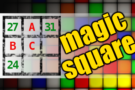

MAGIC SQUARE: Calculate A-B+C
The aim is to place the some numbers from the list (11, 12, 18, 24, 25, 26, 27, 31, 33, 86, 96) into the empty squares and squares marked with A, B an C. Sum of each row and column should be equal. All the numbers of the magic square must be different. Find values for A, B, and C. Solution is A-B+C.Correct answers: 2
#brainteasers #math #magicsquare

A lady noticed her husband sta...
A lady noticed her husband standing on the bathroom scale, sucking in his stomach.
Thinking he was trying to weigh less with this maneuver, she commented, "I don't think that's going to help."
"Sure it does," he said. "It's the only way I can see the numbers."
Thinking he was trying to weigh less with this maneuver, she commented, "I don't think that's going to help."
"Sure it does," he said. "It's the only way I can see the numbers."